Primer Design
Download "Primer Design" Ebook
Primers in DNA Amplification and Scientific Applications
DNA primers have been developed for use in molecular biology methods such as the ubiquitous polymerase chain reaction (PCR). Tips for how to design primers for PCR is the main focus in this article and is discussed in the next section. However, primers are also used in DNA sequencing and recombinant DNA technology methods such as restriction cloning and newer DNA assembly methods such as Golden Gate and Gibson DNA assembly methods — all of which are also offered in Benchling.
Primer Design for Cloning
Molecular cloning is an important technique that allows scientists to replicate specific target segments of DNA in host cells. The target gene or fragment of interest can be inserted into a cloning vector, generally a circular prokaryotic plasmid genome (sometimes simply called a plasmid vector), which can then be transformed and taken up by a specific recipient cell line, usually a bacterial cell. The vector will then be incorporated into the host genome via DNA recombination, and as the cell line naturally undergoes DNA replication as outlined above, the target gene of interest will also replicate, or begin being cloned.
But first scientists must isolate the specific target gene to be cloned. They can do this by using PCR to amplify the target gene if it’s already isolated. Alternatively, if the desired sequence to be amplified is in a plasmid or in another sequence, they can find restriction sites flanking the sequence in order to cut it out using restriction enzymes, or rather, digest the sequence. Both methods allow scientists to produce a specific stretch of DNA that they can then assemble into a plasmid vector to be transformed into cell lines for cloning.
Primer Design for Digest and Ligate Cloning Method
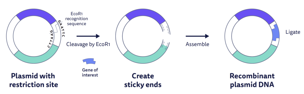
Common restriction enzymes (also called restriction endonucleases) such as EcoR1 recognize and cut at specific palindromic restriction sites in the plasmid. EcoR1 cuts in a zig-zag across the double-stranded DNA creating sticky ends, or single-stranded DNA overhangs of a few base pairs long. Other enzymes like Alu1 are blunt end restriction enzymes that create blunt ends without overhangs. Generally, the same restriction enzymes that cut the plasmid are also used to digest the gene of interest out of its parent DNA, creating complementary sticky ends in the plasmid and target sequence. Once the two are combined in solution, the target fragment can ligate into the plasmid through hydrogen bonding. Gaps are again filled by DNA ligase and the plasmid is ready to be used in further experiments, such as transformation.
This is considered the traditional digest and ligate method that will ultimately create recombinant DNA. Recombinant DNA is formed in the laboratory by combining genetic material from multiple sources, including DNA material from two different species.
Another common alternative is to design primers with restriction sites at their 5’ ends. After PCR, this creates a copy of the target DNA region with restriction sites needed at the end of the fragment. This method is used when there may not be restriction sites for the same enzymes in the correct place in both the vector and insert.
Traditional digest and ligate methods limit scientists to a single insert per cloning cycle. Advances in cloning technologies have focused more on the assembly of multiple DNA fragments, which is made much more efficient with assembly techniques such as Golden Gate cloning and Gibson cloning.
Primer Design for the Golden Gate Cloning Method
Also known as Golden Gate assembly, this cloning method allows scientists to simultaneously assemble a vector (the backbone, usually a plasmid) with one or multiple DNA insert fragments (the parts) into a single construct. Golden Gate uses Type IIs restriction enzymes, which unlike the standard Type II enzymes, cut outside of their recognition sites, creating non-palindromic sticky end overhangs. This allows for multiple fragments of DNA to be assembled by using combinations of overhang sequences on the insert fragments that allow adjacent fragments to easily anneal together. The Type IIs restriction enzymes allow for Golden Gate assembly to be considered scarless, meaning it leaves no restriction sites left over after cloning.
Designing primers for Golden Gate cloning is automatic with Benchling’s primer design and DNA assembly tools. For Golden Gate, the PCR primers should overlap adjacent DNA fragments to include restriction sites and be designed in a way that when digested with a Type IIS enzyme, directional assembly of the fragments is possible with DNA ligase.
Benchling’s Golden Gate Assembly wizard tool was built in conjunction with scientists from New England Biolabs. The assembly wizard helps to automate the time-consuming and error-prone process of selecting appropriate sticky ends, while also supporting more complex techniques like site-directed mutagenesis and the integration of spacers between fragments.
Primer Design for the Gibson Cloning Method
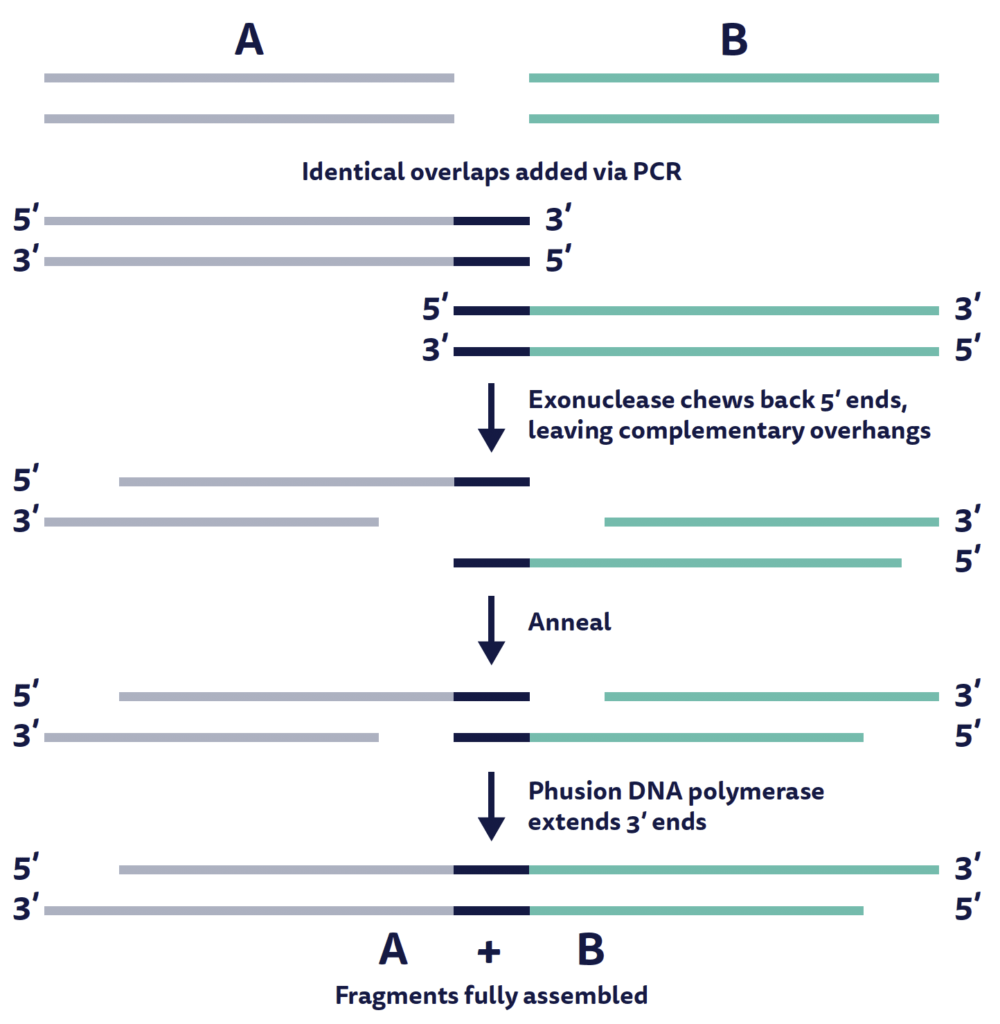
Also known as Gibson assembly and created by Daniel G. Gibson in 1996, the Gibson cloning protocol is similar to the Golden Gate method in that it is scarless and can simultaneously assemble a backbone sequence with multiple DNA fragments into a single construct. What’s unique about Gibson cloning is that it relies on recombination rather than restriction digestion and ligation to generate plasmids.
Gibson assembly requires scientists to produce identical homologous overlaps at the ends ~20-40 base pairs long on both the target DNA fragments to be assembled, and on each side of the linearized vector. This creates an identical overlap that can be chewed back by exonuclease, leaving complementary overhangs to assist in assembly.
Powering breakthroughs for over 1,300 biotechnology companies, from startups to Fortune 500s
